Upper Thigh Anatomy / Like the forearm, the upper leg, or thigh, has a dense arrangement of many muscles.. …front and sides of the thigh. Pelvic & upper thigh anatomy. Upper leg anatomy and function. Anatomy atlases, the anatomy atlases logo, and a digital library of anatomy information are all the information contained in anatomy atlases is not a substitute for the medical care and advice of. The thigh is the area between the hip and the knee joint.
Other articles where thigh is discussed: It's the area that runs from the hip to the. The anatomical areas found on the upper limb can serve as key landmarks to help us find important anatomical structures such as finding one of the superficial veins: •medial thigh muscles•adductor longus muscle•adductor magnus muscle. • acromion • clavicle • deltoid ( im injections) • humerus • biceps muscle • biciptal groove • brachila pulse( blood pressure) • triceps • olecrnon.
How To Fix Upper Thigh Pain When Walking (2020 GUIDE ... from www.arcticdry.co.uk Anterior muscles extend your legs. The thigh is the area between the hip and the knee joint. • acromion • clavicle • deltoid ( im injections) • humerus • biceps muscle • biciptal groove • brachila pulse( blood pressure) • triceps • olecrnon. Start studying thigh/upper leg anatomy. This webpage presents the anatomical structures found on thigh mri. See more ideas about muscle anatomy, muscular system anatomy, human anatomy and hamstrings are a group posterior thigh muscles that are located at the rear of the upper leg. Like the forearm, the upper leg, or thigh, has a dense arrangement of many muscles. Finally, the hamstring muscles that run down the back of the thigh start on the bottom of the pelvis.
The thigh is the area between the hip and the knee joint.
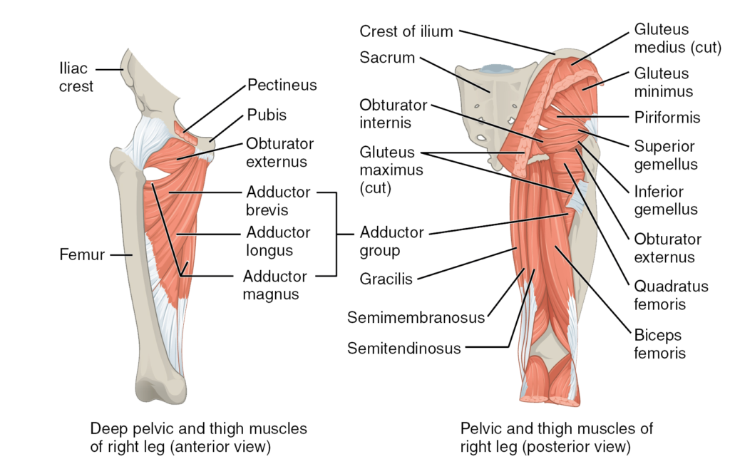
630 anatomical structures of the upper limb (pectoral girdle, shoulder, arm, elbow, forearm, wrist we used the terminologia anatomica to label all the anatomical structures; Upper leg numbness, thigh weakness, thigh pain from overuse. Learn vocabulary, terms and more with flashcards, games and other study tools. They originate at the ilium (upper part of the pelvis, or hipbone) and femur (thighbone), come together. Like the forearm, the upper leg, or thigh, has a dense arrangement of many muscles. This webpage presents the anatomical structures found on thigh mri. In clinical anatomy the thigh muscles are divided into three groups: These images are arranged in radiographic view. Upper part of medial surface of the shaft of tibia. The thigh bears much of the load of the body's weight when a person is upright. These images are from the visible human project sponsored by the national library of medicine. In human anatomy, the thigh is the area between the hip (pelvis) and the knee. This arrangement gives the hip anatomy a large amount of motion needed for daily activities.
Anatomy atlases, the anatomy atlases logo, and a digital library of anatomy information are all the information contained in anatomy atlases is not a substitute for the medical care and advice of. This webpage presents the anatomical structures found on thigh mri. •medial thigh muscles•adductor longus muscle•adductor magnus muscle. Upper part of medial surface of the shaft of tibia. • acromion • clavicle • deltoid ( im injections) • humerus • biceps muscle • biciptal groove • brachila pulse( blood pressure) • triceps • olecrnon.
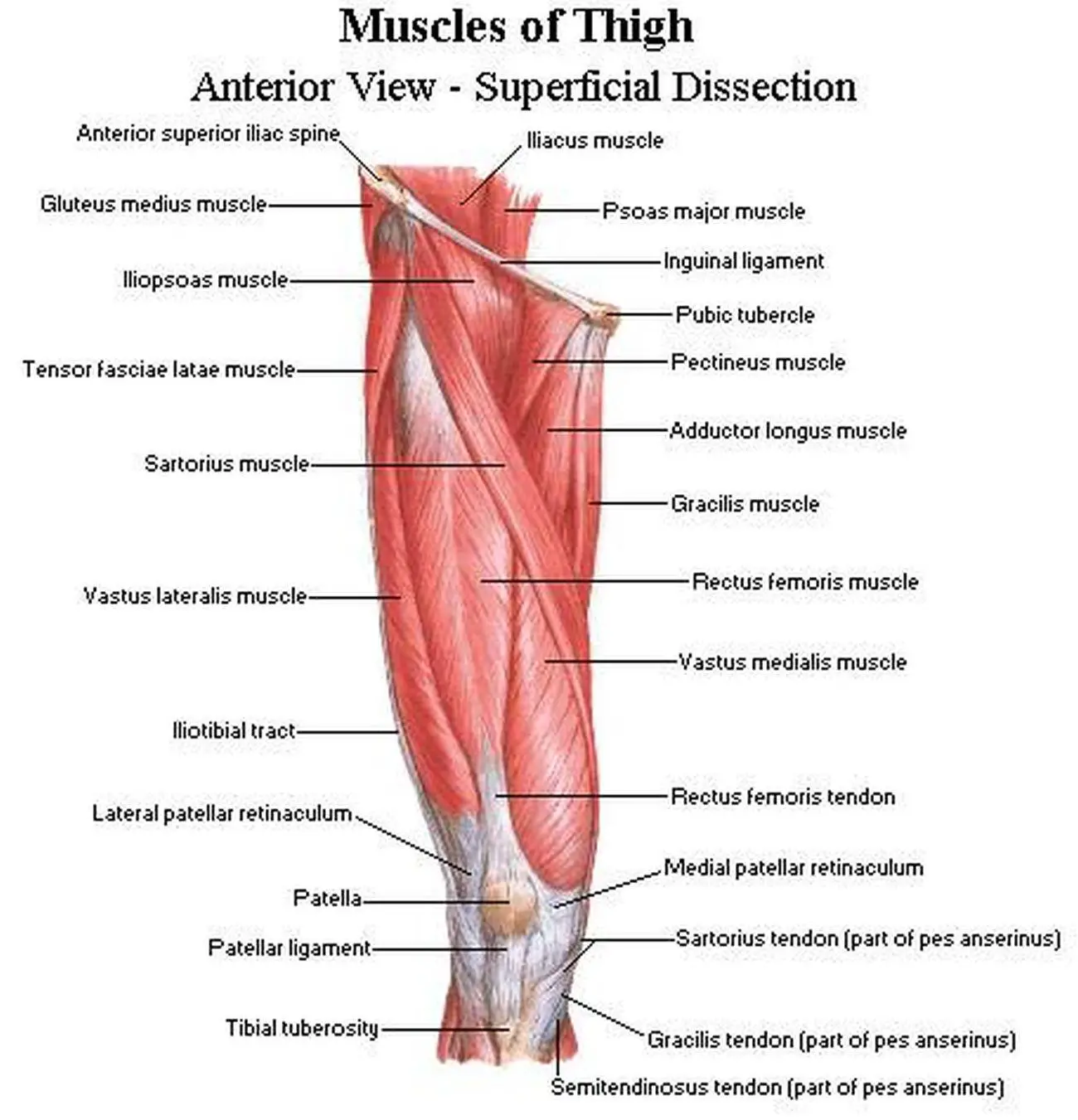
Anterior view of leg muscles from www.anatomynote.com This section of the website will explain large and minute details of arterial anatomy of upper legs (thigh arteries). Ebraheim's educational animated video describes muscle anatomy of the thigh. Pelvic & upper thigh anatomy. The single bone in the thigh is called the femur. Like the forearm, the upper leg, or thigh, has a dense arrangement of many muscles. Anatomy, bony pelvis and lower limb, thigh nerves. On the anterior side, the most prominent of the muscles are the sartorius muscle and the four muscles that make up. Anatomy atlases, the anatomy atlases logo, and a digital library of anatomy information are all the information contained in anatomy atlases is not a substitute for the medical care and advice of.
We think this is the most useful anatomy picture that you need.
Upper part of medial surface of the shaft of tibia. Other articles where thigh is discussed: They originate at the ilium (upper part of the pelvis, or hipbone) and femur (thighbone), come together. Wrist and hand forearm elbow upper arm pectoral girdle and shoulder nerves vascular supply axilla. •medial thigh muscles•adductor longus muscle•adductor magnus muscle. Vascular anatomy of the upper arm. Ebraheim's educational animated video describes muscle anatomy of the thigh. Anatomy, bony pelvis and lower limb, thigh nerves. Like the forearm, the upper leg, or thigh, has a dense arrangement of many muscles. …front and sides of the thigh. These images are arranged in radiographic view. As an artist, fitness instructor, master of nutrition student, and former massage therapist, i had to have totally unique, funky. Anterior muscles extend your legs.
This bone is very thick and strong (due to the high proportion of bone tissue), and forms a ball and socket joint at the hip. Anterior muscles extend your legs. …front and sides of the thigh. Anatomically, it is part of the lower limb. Pain in the upper thighlearn about different causes of upper thigh pain, from injuries to nerve problems.
Pictures Of Anterior Thigh Muscles from healthiack.com …front and sides of the thigh. Finally, the hamstring muscles that run down the back of the thigh start on the bottom of the pelvis. Learn vocabulary, terms and more with flashcards, games and other study tools. In clinical anatomy the thigh muscles are divided into three groups: It's the area that runs from the hip to the. We look at the associated symptoms and treatment options. These images are arranged in radiographic view. Anterior muscles extend your legs.
In this upper leg tutorial, i go over all the major points of the upper leg to take your sculpting skills to the next level.
…front and sides of the thigh. • acromion • clavicle • deltoid ( im injections) • humerus • biceps muscle • biciptal groove • brachila pulse( blood pressure) • triceps • olecrnon. Like the forearm, the upper leg, or thigh, has a dense arrangement of many muscles. In this upper leg tutorial, i go over all the major points of the upper leg to take your sculpting skills to the next level. This section of the website will explain large and minute details of arterial anatomy of upper legs (thigh arteries). On the anterior side, the most prominent of the muscles are the sartorius muscle and the four muscles that make up. Learn vocabulary, terms and more with flashcards, games and other study tools. Start studying thigh/upper leg anatomy. We look at the associated symptoms and treatment options. The single bone in the thigh is called the femur. In clinical anatomy the thigh muscles are divided into three groups: In human anatomy, the thigh is the area between the hip (pelvis) and the knee. •medial thigh muscles•adductor longus muscle•adductor magnus muscle.


0 Comments